
Animal tissues are groups of similar cells that work together for one job, and the body runs on four main types: epithelial, connective, muscle, and nervous. Each type has a different shape and layout, and that design tells you what it can do. Flat cells help cover surfaces. Long cells help contract. Fiber-rich tissue helps hold parts together. Signal-carrying tissue helps the body react fast. That setup matters in an introduction to biology II course because tissues sit right between cells and organs. If you understand tissues, the rest of anatomy starts to make sense instead of looking like a pile of random names. In a college credit biology class, this topic shows up early because it links cell structure to real body jobs. It also fits study online formats well, since you can learn the four tissue types with diagrams, slides, and practice questions. The big idea is simple: structure drives function. A tissue does not look the way it does by accident. A sheet of tight cells can block water. A web of fibers can absorb force. A bundle of elongated cells can shorten and pull. A network of neurons can send signals in milliseconds. Those design rules show up again and again in organs and organ systems, which is why this topic keeps coming back in biology, health science, and pre-med work.
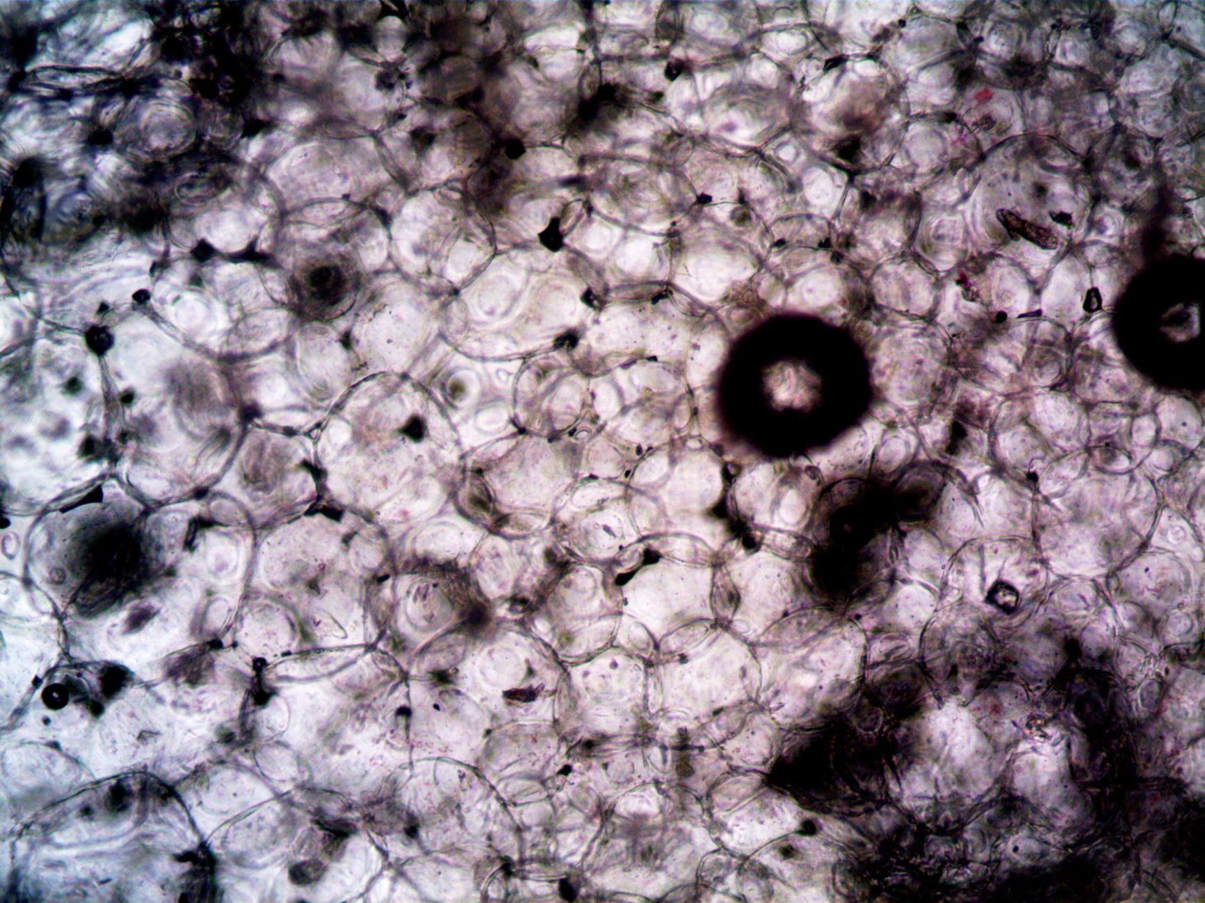
What Are Animal Tissues in Biology?
Animal tissues are groups of similar cells that work together for one shared job, and biology classes usually sort them into 4 main types: epithelial, connective, muscle, and nervous. That idea sits at the center of Introduction to Biology II, because it connects cell shape, organ function, and body systems in one clean model.
Think of tissues as the middle step between single cells and full organs. A cell by itself can do only so much. A sheet of 1000s of epithelial cells can cover a surface. A bundle of muscle cells can move a bone. A network of nerve cells can send signals across the body in less than 1 second.
This topic matters in college-credit biology courses because it shows how living systems stay organized. Skin, stomach, heart, and brain all depend on more than one tissue type, and each tissue brings a different job to the table. That is why a good introduction to biology II course spends time here instead of racing past it.
The catch: If you memorize the names and skip the structure, you miss the whole point. Animal tissues only make sense when you connect cell shape to function, and that is where most students either get it or get lost.
Online learners do well here because tissue diagrams, microscope images, and quiz banks make the patterns easy to spot. A study online format works best when you keep matching examples to the 4 tissue types until the differences stop looking fuzzy.
Why Does Tissue Structure Match Function?
Tissue structure matches function because cell shape, thickness, fibers, and signaling lines decide what a tissue can do, and biology does not waste space on useless design. A 1-cell-thick epithelial layer can move gases fast. A dense connective layer can take stress. A long muscle cell can shorten. A neuron can fire a signal in a fraction of a second.
That rule shows up all over the body. Thin squamous epithelial cells in the lungs help oxygen and carbon dioxide move across a small barrier. The intestinal lining uses a simple columnar shape and tiny surface folds to absorb nutrients over a huge area. Bone uses hard mineral plus collagen fibers, and that mix gives you strength without turning the body into stone. These are not random details. They are the job.
Reality check: A tissue that needs fast exchange cannot be thick and sloppy. A tissue that needs support cannot be fragile. A tissue that needs movement cannot stay short and rigid. That sounds obvious, but a lot of students still treat anatomy like a memory game instead of a design problem.
Muscle tissue gives the best example. Skeletal muscle cells stretch long and line up in bundles so they can pull in one direction. Cardiac muscle cells branch and connect with special junctions so the heart can beat 60 to 100 times per minute without falling out of rhythm. Smooth muscle cells stay spindle-shaped because they need slow, steady squeezing in the stomach, intestines, and blood vessels.
Structure also helps explain failure. Damage the basement membrane under epithelial tissue, and the lining loses support. Break collagen in connective tissue, and joints or skin weaken. Interrupt neuron signaling, and movement or sensation drops fast. That is not a side note. That is biology working the way it was built.
Learn Biology 2 Online for College Credit
This is one topic inside the full Biology 2 course on UPI Study — a self-paced, online class that earns real college credit. Credits are ACE and NCCRS evaluated and transfer to partner colleges across the US and Canada. Courses start at $250 with no deadlines and lifetime access.
See Biology 2 Course →Which Features Define Epithelial Tissue?
Epithelial tissue covers surfaces, lines spaces, and handles protection, absorption, secretion, and filtration. It usually forms thin sheets with tightly packed cells, and that tight packing matters more than fancy vocabulary. A 1-cell-thick layer can exchange fast. A 30-second glance at a skin slide can show why this tissue looks so different from muscle or bone. Worth knowing:
- Epithelial cells sit close together with very little space between them. That tight fit helps skin and gut lining block leaks and germs.
- Most epithelial tissue shows polarity, which means the top side and bottom side do different jobs. The bottom side attaches to a basement membrane.
- Many epithelia turn over fast. Skin cells can renew in about 2 to 4 weeks, which helps the body replace worn-out cells quickly.
- Simple squamous epithelium uses a thin 1-cell layer for diffusion and filtration. You see that design in lung air sacs and kidney filters.
- Simple columnar epithelium lines much of the intestine. Its tall cells help absorb nutrients and release mucus from gland cells.
- Stratified epithelia use multiple layers for protection. Skin, or the epidermis, needs that extra depth because it takes daily wear.
- Gland tissue comes from epithelial cells too. Sweat glands and digestive glands both show how one tissue type can cover, line, and secrete.
How Do Connective, Muscle, and Nervous Tissues Work?
Connective, muscle, and nervous tissues handle support, movement, and communication, and the body fails fast when one of them breaks. Connective tissue uses cells plus an outside matrix, muscle tissue uses contractile proteins, and nervous tissue uses electrical and chemical signals. That split sounds neat on paper, but real organs mix all 3 in messy, useful ways. A heart wall, for instance, needs muscle to pump, connective tissue to hold shape, and nerves to keep timing steady. Bottom line:
- Connective tissue includes bone, cartilage, fat, blood, and loose connective tissue. Its fibers and matrix give support, storage, transport, and repair.
- Collagen fibers matter here. They give tendons and ligaments high strength, and that is why they resist stretching so well.
- Skeletal muscle moves bones under voluntary control. A biceps curl or a 10-minute walk both depend on it.
- Cardiac muscle contracts in the heart wall without a pause button. It keeps blood moving about 24 hours a day.
- Smooth muscle works in the stomach, intestines, and blood vessels. It squeezes slowly, and that steady motion fits its job better than quick bursts.
- Neurons carry signals, and glial cells support them. Together they let the brain, spinal cord, and nerves talk in milliseconds.
Introduction to Biology II usually spends a full unit on these 3 tissue groups because they explain how the body keeps structure, movement, and control in sync. That focus is deserved. These tissues do the heavy lifting, and any course that skips them leaves a hole you will feel later.
How Do Animal Tissues Build Organs?
Organs form when 2 or more tissue types work together, and that is why the stomach, heart, and skin do far more than any single tissue could do alone. The stomach needs epithelial tissue for lining, smooth muscle for churning, connective tissue for support, and nervous tissue for timing. A heart needs cardiac muscle to pump, connective tissue to keep valves and chambers shaped right, and nerves to keep the beat coordinated. Skin uses epithelial tissue for protection, connective tissue for strength, and nerve endings for sensation.
That pattern shows up across organ systems too. The digestive system depends on epithelium for absorption and secretion, muscle for movement, and nerves for control. The circulatory system depends on cardiac muscle, blood as a connective tissue, and nerves that adjust heart rate and vessel tone. You do not get a working organ because one tissue is amazing. You get a working organ because 4 tissue types do different jobs at the same time.
What this means: If one tissue type fails, the whole organ feels it. A 1-millimeter break in lining tissue can cause leakage. Weak connective tissue can make an organ lose support. Bad nerve signaling can throw off rhythm or reflexes. Biology gets practical fast here.
This is why animal tissues show up early in an introduction to biology II course and why they matter in college credit study. Once you see how tissues build organs, anatomy stops looking like a list and starts looking like a system with logic. That shift is the real win, and it carries into every later unit on physiology and homeostasis.
Frequently Asked Questions about Animal Tissues
There are 4 main animal tissue types: epithelial, connective, muscle, and nervous. You see them in every organ, from skin and stomach lining to bone, blood, and nerves, and each one does a different job that the body needs.
The first step is to look at cells that group together and do one job, like lining, support, movement, or signaling. In anatomy and in an introduction to biology ii course, that’s the whole point of tissue: form matches function.
What surprises most students is that epithelial tissue does more than cover the outside of the body. It lines organs like the lungs and intestines, and its tightly packed cells help with protection, absorption, and secretion.
The most common wrong assumption is that connective tissue just means bone. It also includes cartilage, fat, and blood, and its loose or rigid structure lets it support organs, store energy, and move materials through the body.
Most students memorize the 3 muscle types—skeletal, cardiac, and smooth—but what actually works is linking each one to a job. Skeletal muscle moves bones, cardiac muscle pumps the heart, and smooth muscle moves food and blood.
This applies to anyone taking biology, anatomy, or an online course with a lab unit on tissues, and it doesn't help much if you skip the organ examples. If you want college credit or ace nccrs credit, you need the tissue names and functions cold.
If you get nervous tissue wrong, you miss how the body sends signals fast through neurons and supporting glial cells. That mistake hurts later topics too, because nerves control reflexes, sensation, and organ communication across the whole system.
Are animal tissues studied only in advanced classes? No, they show up early in introduction to biology ii and in many study online units because they explain how organs work. The big idea stays the same: structure supports function.
They work together in an organ by each doing a separate job: epithelial tissue lines surfaces, connective tissue supports, muscle tissue moves, and nervous tissue sends signals. In the stomach, those 4 tissue types help digestion happen in order.
Yes, animal tissue units can count toward transferable credit in courses that award accepted biology credit, especially when the course uses recognized standards like ACE and NCCRS. You still need the tissue content, because exams often test structure, function, and organ examples in 1 unit.
Final Thoughts on Animal Tissues
Animal tissues are not just four words to memorize. They are the body’s working parts, and each one earns its place by doing a different job. Epithelial tissue covers and lines. Connective tissue supports and binds. Muscle tissue moves. Nervous tissue sends messages. That lineup sounds simple, but the simple version hides the real beauty: structure and function always move together. Once you start reading tissues as design, organs make more sense. The stomach needs a lining, force, and control. The heart needs pumping tissue, support tissue, and signal timing. Skin needs a barrier, a frame, and sensation. None of those jobs works alone. Biology does not build with single bricks. It builds with teams. That is why this topic shows up so often in intro biology, anatomy, and health science. You keep using it because it keeps paying off. A good tissue chart can help, but real understanding comes from comparing shapes, layers, fibers, and signal paths until the pattern sticks. Then the names stop feeling random. If you are studying this now, focus on 2 questions for every tissue: What does it look like, and what job does that shape let it do? Answer those two things, and you will remember far more than a list of terms. Next, review one organ at a time and match each tissue to its role.
How UPI Study credits actually work
Ready to Earn College Credit?
ACE & NCCRS approved · Self-paced · Transfer to colleges · $250/course or $99/month