
The cytoskeleton in biology is the cell’s internal support network, and it does far more than hold shape. It gives a cell structure, helps it move, moves cargo inside the cell, and helps it split into two new cells during division. If you think of a cell as a tiny living factory, the cytoskeleton acts like beams, roads, and cables all at once. Students in an Intro to Biology I course meet this topic early because it shows how cells stay organized instead of floating around as blobs of fluid. That matters in real life. A nerve cell can stretch long distances, a muscle cell can contract with force, and an immune cell can crawl toward a threat. Each of those jobs depends on the cytoskeleton. Three parts do most of the work: microfilaments, intermediate filaments, and microtubules. Each one has a different size and job, and that split matters. Microfilaments help with shape and movement near the membrane. Intermediate filaments give cells toughness. Microtubules act like track lines for transport and the spindle in mitosis. Once you see those parts working together, the cell stops looking random and starts looking engineered.
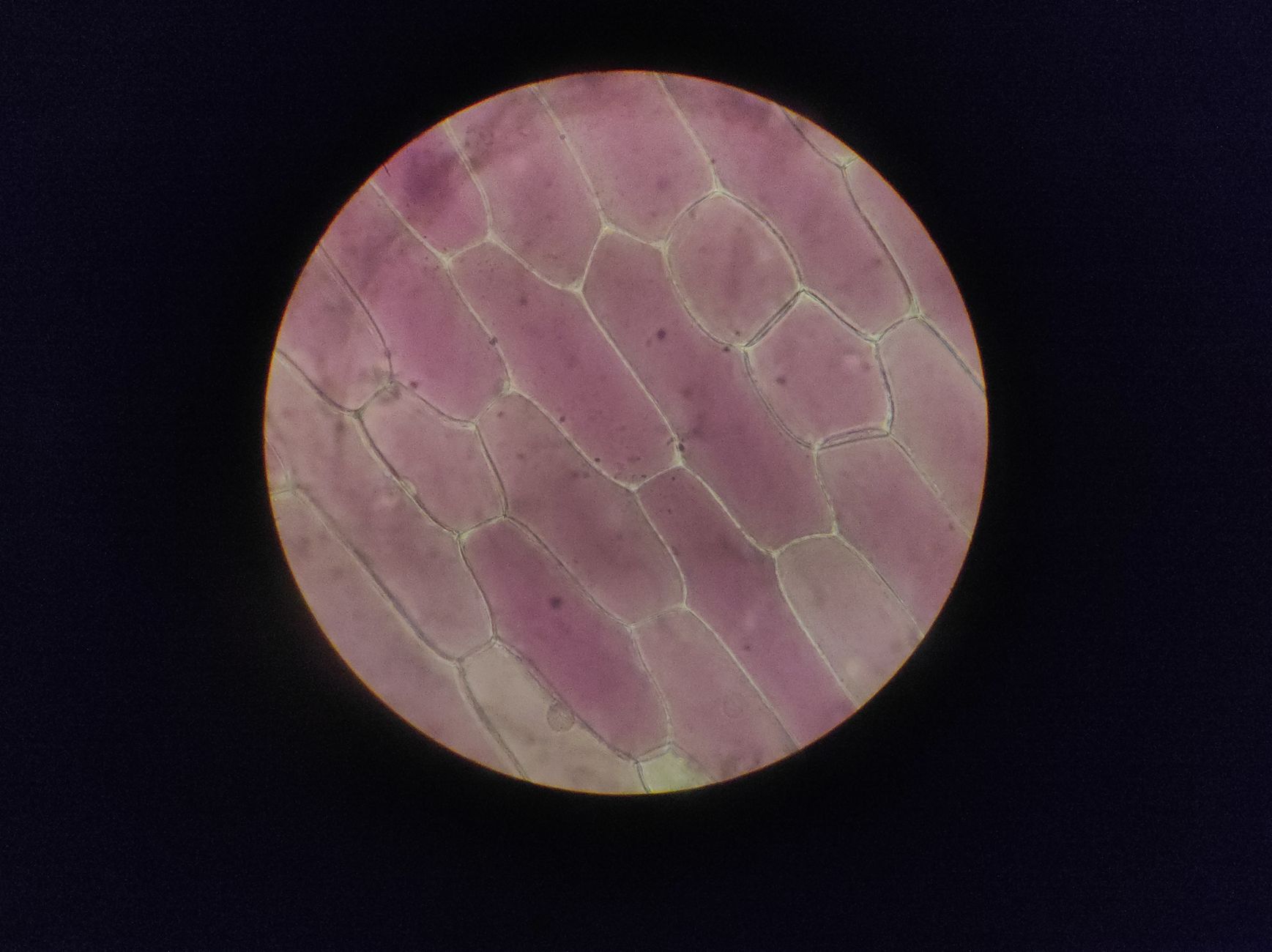
Why Is the Cytoskeleton Essential?
The cytoskeleton is the cell’s internal support network, and it gives a living cell shape, strength, and order in the same way a frame gives a building structure. In an Intro to Biology I course, this idea shows up fast because a cell without internal support would not hold a stable form, move well, or keep its parts in the right place.
The catch: Cells are not static bags of fluid. They change shape, pull in nutrients, and shift their parts across distances as small as 1 micrometer and as large as several cell widths, so the cytoskeleton has to stay active all the time. That constant work matters in nerve cells, muscle cells, and white blood cells, which all depend on a strong but flexible internal framework.
The cytoskeleton also helps cells respond to what happens outside them. A cell can stretch, tighten, crawl, or divide because protein fibers inside it can grow, shrink, and rearrange in minutes. That is a lot more interesting than a passive support scaffold, and it is the part students should remember first. Shape alone would already make this system important, but the cytoskeleton does more than keep a round cell round or a flat cell flat.
It also organizes the inside of the cell. Organelles do not just drift aimlessly if the cell needs order. A cell can place a nucleus, send vesicles, and move proteins to exact spots because the cytoskeleton gives the cell a working layout. In a biology lab, that is the difference between a tidy, functional cell and a chaotic one that cannot keep up with a 24-hour life cycle.
What Are Microfilaments in the Cytoskeleton?
Microfilaments are the thinnest fibers in the cytoskeleton, and they are built mainly from actin, a protein that can form long chains only about 7 nanometers wide. That tiny size hides a big job: these filaments support cell shape, help cells change form, and power movement near the membrane.
The cell cortex sits just under the plasma membrane, and microfilaments crowd that zone like a tight ring of scaffolding. Worth knowing: That thin shell matters because it lets the cell push out fingerlike projections, squeeze through narrow spaces, and change shape fast during growth or motion. A crawling immune cell uses actin-based changes to move across tissue, and a dividing animal cell uses actin to help pinch itself in two.
Muscle contraction also depends on actin. In skeletal muscle, actin works with myosin so the fibers can slide past each other and shorten a cell by a measurable amount. That same sliding idea appears in cell movement too, which is one reason biology teachers keep returning to actin in different chapters. It shows up in structure, transport, and motion, not just one neat box.
Microfilaments can feel abstract at first, but they are the reason a cell can bend without breaking. That flexibility has a downside too: if actin control fails, a cell loses shape and motion fast. Students who want clear course notes often pair this topic with Introduction to Biology I because actin comes up again and again in cell biology.
Learn Biology 1 Online for College Credit
This is one topic inside the full Biology 1 course on UPI Study — a self-paced, online class that earns real college credit. Credits are ACE and NCCRS evaluated and transfer to partner colleges across the US and Canada. Courses start at $250 with no deadlines and lifetime access.
Explore Biology 1 Course →How Do Intermediate Filaments Support Cells?
Intermediate filaments give cells toughness, and they do it without trying to move the cell or act like tracks. Their fibers sit between microfilaments and microtubules in size, usually about 10 nanometers across, which makes them a sturdy middle layer in the cytoskeleton.
Reality check: Cells face stretching, pulling, and shear forces all day, especially in skin, muscle, and organ linings, so they need a system that resists damage instead of just changing shape. Intermediate filaments do that job well. They help cells hold together under stress, and they make tissues like skin and the lining of the gut much harder to tear.
These filaments also support cell architecture inside the nucleus and the cytoplasm. Nuclear lamins, a type of intermediate filament, line the inside of the nuclear envelope and help keep the nucleus stable during the cell cycle. That detail shows up in college biology courses because it connects cell shape to gene control and division.
This part of the cytoskeleton can seem less flashy than movement or transport, but I think it may be the most underrated piece. Without it, cells would bend too easily and tissues would fail under pressure. Students often remember actin and microtubules first, then forget the “middle” class of fibers, which is a mistake. Durable cells need more than motion. They need resistance. Introduction to Biology I usually covers that link when it reaches cell structure and tissue function.
How Do Microtubules Organize Cell Transport?
Microtubules are the largest cytoskeletal fibers, and they are built from tubulin into hollow tubes about 25 nanometers wide. That size lets them act like strong internal tracks, which is why cells use them to move vesicles, position organelles, and build the spindle apparatus during division. A cell that needs to send a cargo packet 10 micrometers across the cytoplasm does not want chaos; it wants a direct route, and microtubules give it one. They also help a cell keep its overall shape, especially in cells that stretch or change position during growth and transport.
Bottom line: Microtubules turn the inside of the cell into a managed system, not a random soup. That is the part students tend to miss on a first pass.
- Vesicles ride microtubules across the cytoplasm using motor proteins like kinesin and dynein.
- Organelle positions stay organized, with mitochondria and the Golgi apparatus placed where the cell needs them.
- The spindle apparatus forms from microtubules during mitosis and lines up chromosomes at the cell’s center.
- Some microtubules grow and shrink fast, which helps cells change shape in seconds to minutes.
A student taking Introduction to Biology I usually meets microtubules when the course shifts from structure to transport, and that is a smart teaching move because the same fibers do both jobs. If you want a second science course that supports this topic, Chemistry I helps with protein structure and motor function. Microtubules can fail in ugly ways too; when they do, transport slows and division goes wrong fast.
How Does the Cytoskeleton Help Cell Division?
The cytoskeleton drives cell division by reshaping itself during mitosis and cytokinesis, and that process depends on microtubules, actin, and careful timing across 4 main phases. DNA copies matter, but the cell still has to move chromosomes, split the cytoplasm, and build two working daughter cells.
During mitosis, microtubules form the mitotic spindle, which attaches to chromosomes through their kinetochores and pulls sister chromatids apart. That action keeps each new cell from getting the wrong set of chromosomes, and it happens in a sequence students usually track as prophase, metaphase, anaphase, and telophase. The spindle is not decoration. It is the machinery.
Cytokinesis finishes the job. In animal cells, an actin-based contractile ring tightens around the cell’s middle like a drawstring, and that squeeze splits one cell into 2. Plant cells use a different setup because their rigid wall changes the process, which makes this topic a nice reminder that cell type matters. A cell that cannot manage this choreography ends up with broken copies or extra nuclei, and that can wreck tissue growth.
The cytoskeleton makes division possible because it gives the cell force, direction, and timing at the same time. That is the part worth remembering for an exam and for real biology. A cell does not divide by DNA alone; it divides because its internal fibers know how to move, pull, and pinch on cue.
Frequently Asked Questions about Cytoskeleton
Most students memorize the parts first, but what works better is to picture the cytoskeleton as the cell’s support frame and road system. It has 3 main parts: microfilaments, intermediate filaments, and microtubules, and they help the cell keep its shape, move, and split during cell division.
This applies to anyone studying cell biology in high school, intro to biology i, or an intro to biology i course, and it does not apply only to advanced lab students. Every eukaryotic cell uses the cytoskeleton, from plant cells to animal cells, because shape, transport, and division all depend on it.
3 main parts make up the cytoskeleton: microfilaments, intermediate filaments, and microtubules. Microfilaments use actin, microtubules use tubulin, and intermediate filaments give extra strength, so each part handles a different job inside the cell.
If you get this wrong, you usually mix up structure with function and lose easy points on cell diagrams, mitosis questions, and transport questions. A common mistake is saying the cytoskeleton only gives shape, when it also helps organelles move and helps chromosomes separate during division.
The most common wrong assumption is that the cytoskeleton is a dead scaffold like a wall. It actually changes all the time, especially during cell movement, vesicle transport, and mitosis, so it acts more like a living support network than a fixed frame.
Start by matching each part to one job: microfilaments for cell movement, intermediate filaments for strength, and microtubules for transport and cell division. If you can label those 3 roles in 1 minute, you already have the core idea.
No, the cytoskeleton in biology does much more than hold shape. It also moves materials inside the cell, helps cells crawl, and forms the spindle that moves chromosomes apart in mitosis, which is why it matters in both structure and function.
What surprises most students is that the cytoskeleton is always changing, not staying fixed. Microtubules can grow and shrink fast, and that constant change helps the cell react, divide, and move parts around in real time.
Microfilaments are thin actin strands that help the cell move and change shape. They matter most in cell crawling, muscle contraction, and the pinch that splits one cell into 2 during cytokinesis.
Microtubules act like tracks for moving vesicles, organelles, and proteins across the cell. Motor proteins such as kinesin and dynein carry cargo along them, and that transport keeps long cells and busy cells working.
Intermediate filaments give cells strength and help them resist pulling and stretching. They support the nucleus, hold tissues together, and matter a lot in cells that face stress, like skin cells.
The cytoskeleton helps cell division by building the spindle, a microtubule structure that pulls chromosomes apart in mitosis. Without that spindle, the cell can't split DNA evenly into 2 new cells.
Yes, you can study online through a biology online course that offers college credit, ace nccrs credit, and transferable credit at cooperating schools. That matters if you want formal credit from an online course without sitting in a campus lab every week.
Final Thoughts on Cytoskeleton
The cytoskeleton looks small under a microscope, but it controls some of the biggest jobs in cell life. It gives shape, holds parts in place, powers movement, moves cargo, and helps cells divide without falling apart. That mix of structure and motion is why biology students keep seeing it in chapter after chapter. Microfilaments, intermediate filaments, and microtubules each carry their own load. Actin helps cells change shape and crawl. Intermediate filaments handle stress and stretching. Microtubules move vesicles, position organelles, and build the spindle in mitosis. Once you separate those jobs, the whole topic gets easier to study and a lot easier to remember. This is also one of those ideas that shows up everywhere in cell biology, tissue biology, and genetics. A cell does not work because it sits there. It works because its internal fibers keep adjusting to what the cell needs right now. That is a simple idea, but it explains a lot. If you are studying for an exam, focus on the 3 fiber types, their sizes, and their jobs. If you can connect those parts to shape, transport, and division, you already have the core of the topic. Review the diagrams, trace the arrows, and test yourself on what each fiber does next.
How UPI Study credits actually work
Ready to Earn College Credit?
ACE & NCCRS approved · Self-paced · Transfer to colleges · $250/course or $99/month